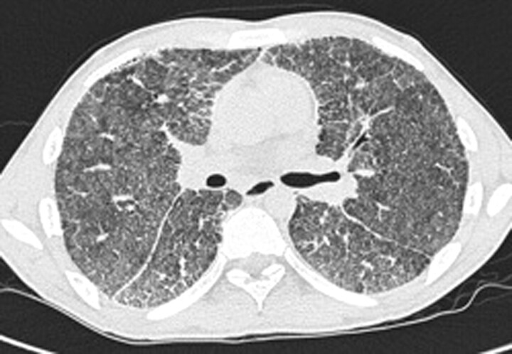
Figure 3.
Case 2. High-resolution computed tomography of the lungs showing bilateral calcifications with increased attenuation involving alveoli, intra- and interlobular septa, fissures and pleura. Signs of fibrosis are visible. Hounsfield unit measurements were very high, representing areas with calcifications.

