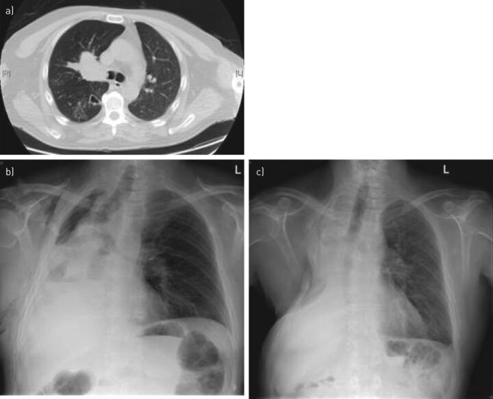
Figure 3.
Right sleeve pneumonectomy for T4 squamous cell carcinoma. a) Chest computed tomography (CT) showing squamous cell carcinoma infiltrating the right main bronchus and carina, with marked obstructive changes and bronchiectasis in the right lower lobe. The right pulmonary artery was infiltrated as well. Cervical mediastinoscopy was performed and showed no invasion of lymph node stations 2R, 4R, 3 and 7. Positron emission tomography-CT showed no distant metastases. The patient was offered extended right pneumonectomy. He developed bronchopleural fistula and had an open pleural window 6 weeks post-operatively. b). He underwent reconstructive thoracoplasty at 2 years and remains disease-free at 4 years, with secondary pulmonary hypertension (c).

