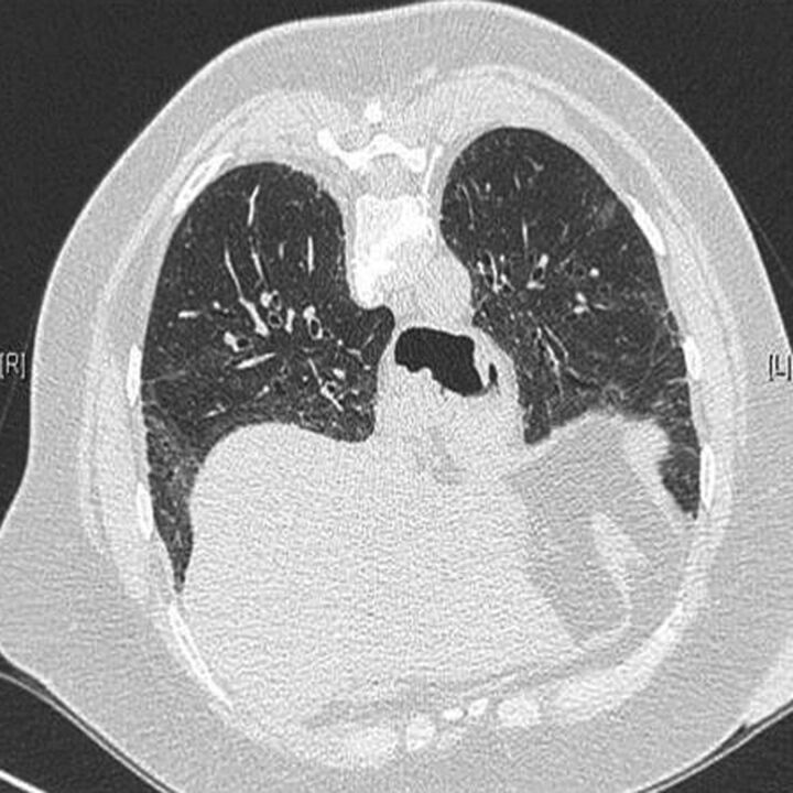
Figure 1.
High-resolution computed tomography scan of a 59-year-old female patient with undifferentiated connective tissue disease, showing patterns of nonspecific interstitial pneumonia (with ground-glass opacification and traction bronchiectasis) and oesophageal dilation. Features of systemic sclerosis were present (Raynaud's phenomenon, megacapillaries at nailfold capillaroscopy, oesophageal hypomotility, anti-topoisomerase-1 antibodies), but no sclerodactily or telangiectasia was visible at 5 years follow-up.

