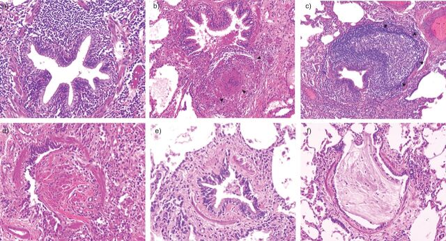
Figure 1.
Representative photomicrographs of individual bronchiolar lesions observed in surgical lung biopsy in patients with small airways disease. a) Cellular bronchiolitis: a narrowed and contracted airway is infiltrated by numerous inflammatory cells without a specific pattern. b) Granulomatous bronchiolitis: the small airway is surrounded by an inflammatory infiltrate with a sarcoid granuloma (arrowheads), which increases the volume of the airway wall resulting in lumen narrowing. c) Follicular bronchiolitis: the small airway is surrounded by a large lymphoid follicule (arrowheads), which increases the volume of the airway wall resulting in lumen narrowing. d) Bronchiolitis obliterans is characterised by lumen obstruction with a fibro-inflammatory polyp. e) Obliterative (constrictive) bronchiolitis: the airways lumen is narrowed by subepithelial fibrosis. Although inflammatory cells and mucous exudates are present within the lumen, no fibro-inflammatory polyp is found. f) Mucous plugging: the airway lumen is obstructed by mucus exudates.

