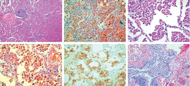
Figure 2.
a–c) This lung biopsy from a 55-year-old male patient, who was a nonsmoker, shows a typical desquamative interstitial pneumonia pattern. Tissue sections show a) diffuse involvement of the lung by intense macrophage accumulations within almost all of the distal airspace, b) without significant thickening of alveolar septa and sparse inflammatory infiltrate (*). Macrophages do not contain dusty pigment. c) Immunohistochemical analysis with CD163 antibody shows the macrophage nature of the cells. d–f) Lung biopsy from a 66-year-old female, who was a heavy smoker, showing a respiratory bronchiolitis/interstitial lung disease pattern. d) Macrophage accumulation is diffuse but predominantly peribronchiolar and f) in respiratory bronchioles. Alveolar septa are slightly thickened. e) The cytoplasm of most macrophages contains an abundant dust-brown pigment.

