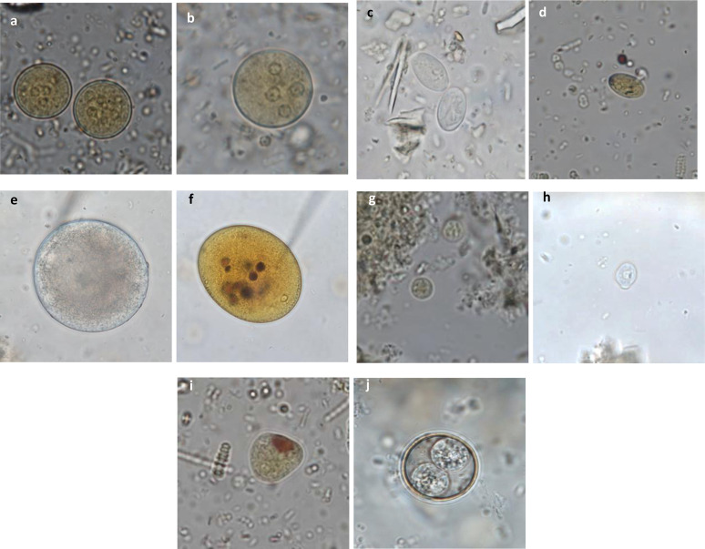
Figure 4.
Diagnostic atlas for the identification of protozoan cysts in non-human primates. (a) Entamoeba sp. cysts stained with Lugol (×100): eccentric nuclei with a voluminous endosome and granulomatous peripheral chromatin; (b) Entamoeba sp. cyst stained with Lugol (×100); (c) Giardia intestinalis cysts (×100): oval cysts of 8–12 μm with a thin outer membrane and 2–4 nuclei and a flagellum; (d) Giardia intestinalis cyst stained with Lugol (×100); (e) Balantioides coli cyst (×100): spherical to ovoid cyst of 50–70 μm in diameter with a thick membrane, granular content, one macro- and one micronucleus; (f) Balantioides coli stained with Lugol (×100); (g) Endolimax sp. cyst stained with Lugol (×100): small round cysts with a thin outer membrane and 1–4 punctiform nuclei with voluminous, irregular endosomes without perisomes; (h) Chilomastix sp. cyst (×100): small piriform cysts with a thin and refringent membrane, one nucleus and a cytostome containing the flagellum; (i) Iodamoeba sp. cyst stained with Lugol (×100): oval cysts with a small round nucleus containing a large vacuole, and a peripheral iodophilic voluminous vacuole; (j) Isospora sp. sporulated oocyst (×100): round cysts containing 2 sporocysts with 4 sporozoites each.

