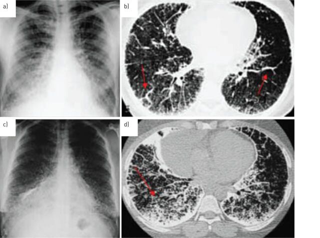
FIGURE 3.
Evolutionary radiological phases of pulmonary alveolar microlithiasis. a) Chest radiograph of the third phase: extremely dense, uniform distribution of radio-opaque bright punctiform elements, with sandstorm appearance. It is not possible to distinguish heart and diaphragm borders in the medial and inferior fields. b) Chest high-resolution computed tomography (HRCT) scan of the third phase: initial calcification of the pleural lining and interlobular septa (arrows), which partly masks the micronodules. c) Chest radiograph of the fourth phase: diffuse microliths with some calcific agglomerates. d) HRCT of the fourth phase: microliths are increased in number, forming calcific agglomerates in some areas, together with intense involvement of the pleural lining and interlobular septa.

