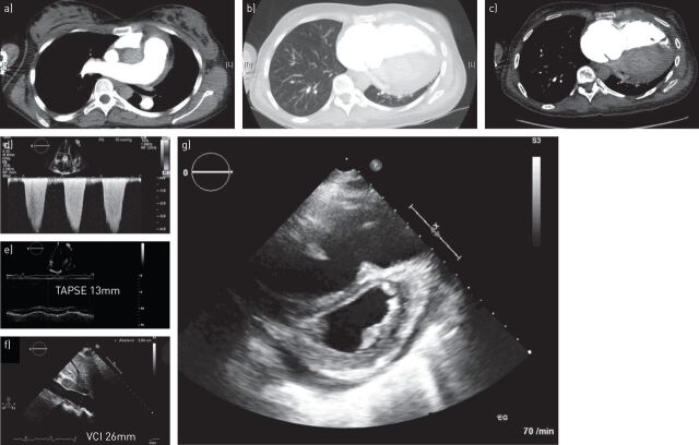
FIGURE 1.
a–c) Chest computed tomography excluding acute lung embolism and parenchymal lung disease and d–g) echocardiography performed during diagnostic work-up in a patient with pulmonary arterial hypertension with right heart failure and active systemic lupus erythematosus (case study 1). d) Elevated tricuspid regurgitant jet velocity; e) reduced TAPSE; f) dilated VCI. TAPSE: tricuspid annular plane systolic excursion; VCI: vena cava inferior.

