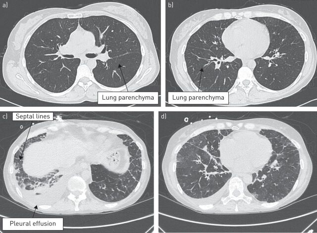
FIGURE 2.
High-resolution computed tomography scans during diagnostic evaluation in a patient with pulmonary veno-occlusive disease, which had been previously diagnosed as idiopathic pulmonary arterial hypertension, showing no evidence of parenchymal disease in a, b) two different slices and c, d) illustrating diffuse ground-glass opacities with thickening interlobular septa (case study 2).

