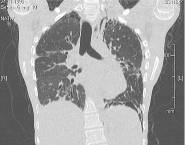
FIGURE 2.
Chest computed tomography scan of a patient with ataxia telangiectasia showing diffuse bibasilar interstitial and interlobular reticular opacities with interlobular septal thickening, and bronchiectasis in the right lower lobe. Note that any radiological imaging should use the minimum possible doses, and concerns about radiosensitivity need to be addressed.

