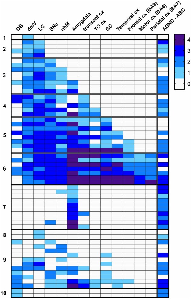
FIGURE 1.

Regional distribution of Lewy pathology in 58 ET cases, with 10-category classification. Severity of Lewy pathology changes is shown by shading, with ordinal gradations from 0 (white) to 4 (darkest shading). ADNC-ABC scores of 0 (white) to 3 (dark shading) scale, corresponding to none, low, intermediate, and high categories. BA, Brodmann area; cx, cortex; dmV, dorsal vagal nucleus; GC, gyrus cinguli; LC, locus coeruleus; nbm, nucleus basalis of Meynert; OB, olfactory bulb; Olf, olfactory; SNc, substantia nigra pars compacta; TO cx, temporal-occipital cortex; transent, transentorhinal.
