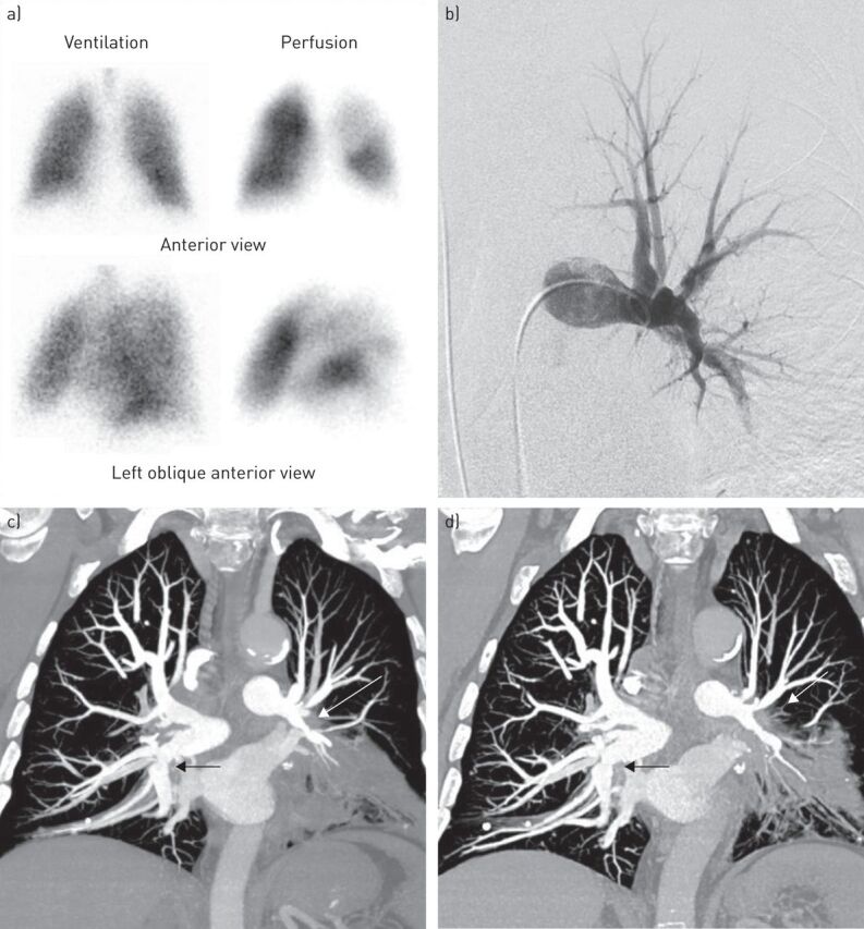
FIGURE 1.

a) Ventilation/perfusion lung scan showing multiple mismatched perfusion defects in the left lower lobe and the left upper lobe. b) Conventional pulmonary angiography. The left anterior view shows an extrinsic compression of the left pulmonary artery. c, d) Contrast-enhanced high-resolution computed tomography of the chest with pulmonary arteries reformatted with maximal-intensity-projection in coronal views. c) The coronal view at baseline shows a right and left mediastinal and hilar soft tissue attenuation mass causing an encasement of the proximal right and left pulmonary arteries (arrows). d) The coronal view after 3 months of corticosteroids shows a mild decrease in the extrinsic compression of the right pulmonary artery whereas the stenosis of the left pulmonary artery remains unchanged (arrows).
