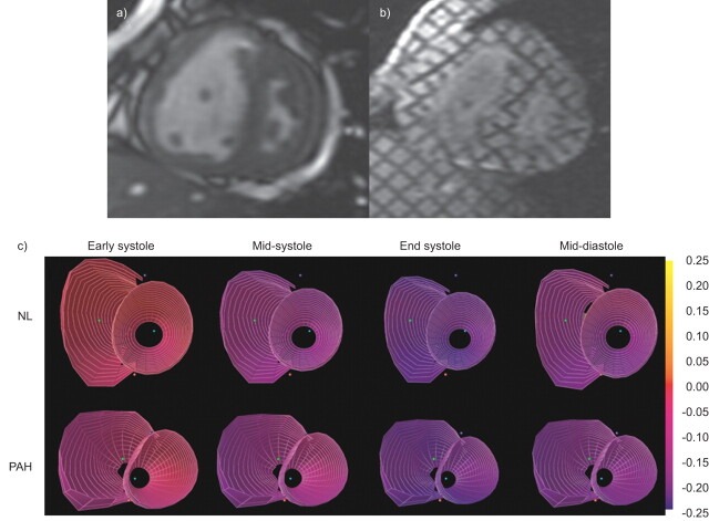
Figure 3.
Cardiac magnetic resonance imaging (MRI) evaluation of cardiac remodelling in pulmonary arterial hypertension (PAH). a) Short axis view of cine-MRI frame at systole and b) corresponding tagged MRI images are shown. c) Three-dimensional strain maps from normal (NL) compared with PAH subjects are shown in various phases of the cardiac cycle. Adapted from [39].

