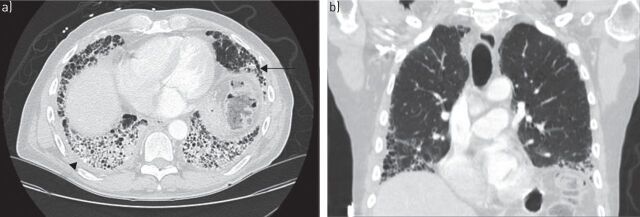
FIGURE 2.
a) Axial and b) coronal computed tomography scans of usual interstitial pneumonia pattern in a patient with rheumatoid arthritis. Subpleural and basilar predominant reticulations, minimal ground-glass opacities, honeycombing (arrow) and pleural thickening (arrowhead) are visible, as well as traction bronchiectasis.

