
Figure 6.
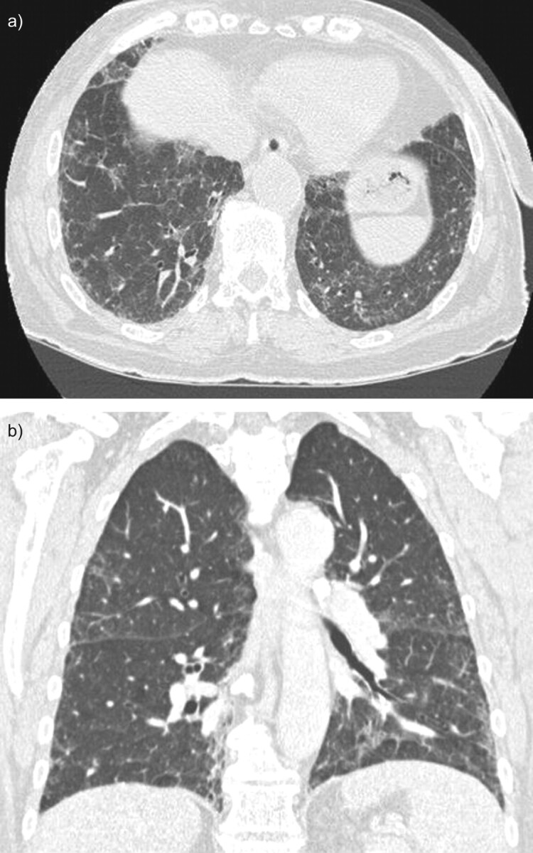
Chest computed tomography (CT) scan of a patient with pulmonary alveolar proteinosis secondary to myelodysplastic syndrome. The CT scan shows ground-glass opacities and reticulations mainly in the lower zone with patchy, slightly peripheral distribution. a) A horizontal section and b) coronal section.