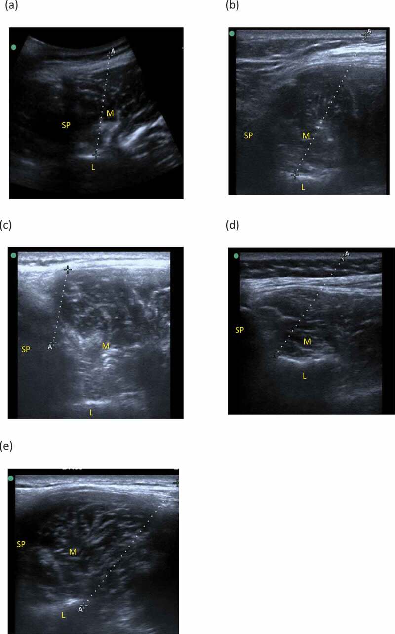
Figure 2.

Ultrasound images of needle placement (the dotted line) in the lumbar multifidus muscle (M): (a) needle reached the lumbar lamina (L) using method 1, (b) needle reached the lumbar lamina (L) using method 2, (c) needle reached the spinous process (SP), (d) needle reached the junction of the spinous process (SP) of the lumbar lamina, (e) needle did not reach a bony landmark. ”A” and ”+” indicate the both ends of the subcutaneous part of the needle.
