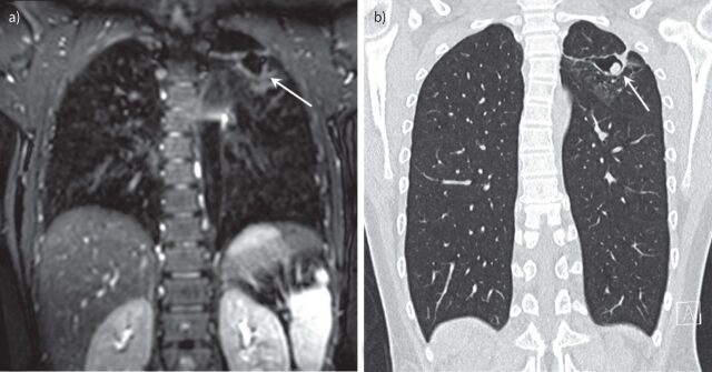
FIGURE 3.
Chest imaging of a 13 -year-old boy with cystic fibrosis. a) Magnetic resonance imaging (MRI) scan (isotropic T2-weighted CUBE, coronal reformat) and b) computed tomography (CT) scan (isotropic coronal reformat) performed 10 months after the MRI. Note cavitary lesion in the left upper lobe on MRI (arrow) with wall thickening but without a solid component. On the follow-up CT, there is a new round solid lesion within the cavity, representing an aspergilloma (arrow) not completely filling the cavity (Modod sign).

