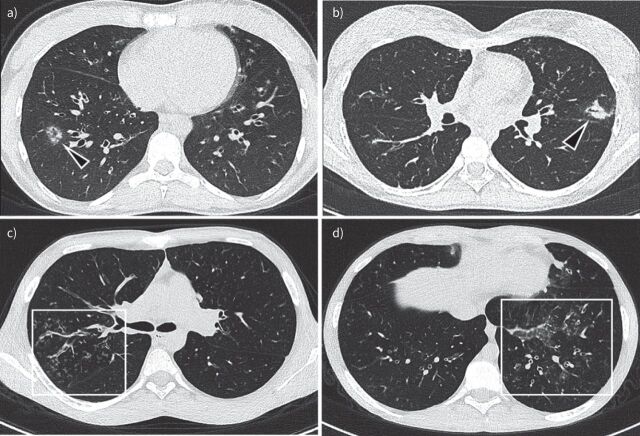
FIGURE 4.
Aspergillus bronchitis/bronchiolitis example images: a) 18-year-old male non-cystic fibrosis (CF) patient, but with sickle cell disease. Multiple nodules with ground glass opacity around peripheral vessels are shown in the right lower lobe (arrowhead in a). b) Chest computed tomography (CT) image of a 17-year-old girl with CF shows Aspergillus bronchopneumonia. Note bronchocentric peri-bronchiolar consolidation on left upper lobe; c and d) Chest CT images of a 15-year-old boy with CF show Aspergillus bronchiolitis. Note filling of peripheral airways with a tree-in-bud pattern and bronchial wall thickening (box in c). In d there is also an area of ground glass (box in d).

