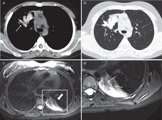
FIGURE 5.
Allergic bronchopulmonary aspergillosis example images. a) and b) Chest computed tomography (CT) images of a 12-year-old boy with cystic fibrosis (CF). a) 2.5 mm axial CT soft tissue window and b) 1 mm axial CT lung window at the same level. Note in a), central bronchiectasis filled with high density material (arrow). In b), note mucus plugs with a typical cylindrical shape originating from the hilar region and extending towards the periphery (arrow) showing typical higher density than paraspinal muscles. c) and d) Lung magnetic resonance imaging scans (T2-weighted axial PROPELLER, in plane resolution 1×1 mm, slice thickness 5 mm) of a 15-year-old girl with CF. c) Atelectasis in the left lower lobe (thick arrow). d) Hypo-intense material filling the central airways (arrows).

