FIGURE 6.

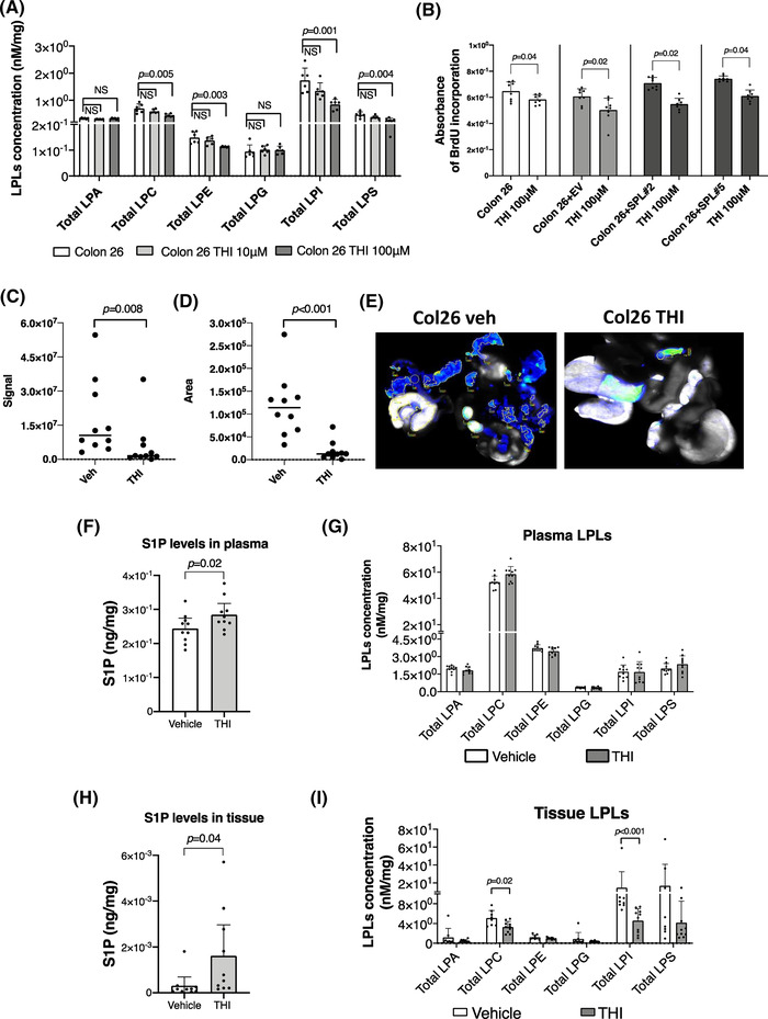
The THI, an inhibitor of the S1P lyase (SPL), worked identical to genetic application both in vitro and in vivo. (A) The dose‐dependent effects of the THI on total levels of the lysophosphatidylcholine (LPC), lysophosphatidylethanolamine (LPE), lysophosphatidylinositol (LPI) in Colon 26 cells. Cells were cultured with 10 μM and 100 μM THI for 72 h and cell lysates including control cell lysates were processed for the measurements of the glyceroLPLs. (B) THI inhibits cell proliferation rates, independently from SPL expression levels. A significant decrease in cell proliferation was observed in cell lines with the addition of the 100 μM THI. Signal (C) and area (D) of the disseminated peritoneal cancer were analyzed in vehicle or THI administered mice by using Odyssey CLx Imaging system. Identical illumination settings (channels: 700/800, resolution: 42 μM, intensities: auto, quality: lowest, analysis: small animal, and focus: 2.0 mm) were used for acquiring all images. (E) Representative scanned images from two groups. Images were acquired and analysed using Image Studio Ver. 4.0 software. (F and H) S1P levels in plasma (F) or disseminated peritoneal cancer tissues (H). S1P levels were significantly increased both in plasma and cancer tissues in THI administered group in comparison to vehicle. (G and I) Total glycero‐LPLs were measured in plasma (G) and disseminated peritoneal cancer tissues (I). In cancer tissues, total LPC and LPI were significantly decreased as an effect of the THI administration. Differences between the two groups were assessed using the unpaired Student's t‐test, while differences among more than two groups were assessed using a one‐way ANOVA. EV, empty vector transfected control cell line; SPL, SPL overexpressing cell line
