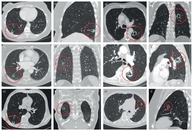
FIGURE 6.
Multiple (type 3) cyst-related primary lung malignancies presenting as cystic airspaces with asymmetrical or circumferential wall thickening. a, b) A 52-year-old female with a biopsy-proven second primary adenocarcinoma in the right lower lobe, synchronous with a T1cN0M1b adenocarcinoma in the left upper lobe (not shown). c, d) A 68-year-old female with a history of breast carcinoma and left upper lobe small cell lung carcinoma. The cyst-related lesion in the right lower lobe was present at baseline and repeatedly misinterpreted as a cavitating contralateral metastasis. After 2 years of continuous increase in wall thickness pathology was obtained and showed a second primary adenocarcinoma. e, f) A 71-year-old male with a history of bladder carcinoma. Cyst-related lesion with progressive circumferential wall thickening on serial computed tomography imaging. The patient opted for stereotactic radiotherapy (STR) without obtaining pathology. g, h) A 78-year-old male without prior malignancy who presented with liver metastasis. This proved to be a moderately differentiated mucinous adenocarcinoma, probably from the lung. Work-up showed this cyst-related lung cancer in the left lower lobe. i, j) An 89-year-old male without a relevant history. Cyst-related lesion in the left lower lobe which showed slight positron emission tomography activity and increase in wall thickness over a period of 3 months. All cultures were negative. STR was performed without obtaining pathology. k, l) A 62-year-old female with a history of an adenocarcinoma in situ in the left upper lobe, treated with radiotherapy. During follow-up this cyst-related lesion developed in the left upper lobe. After consecutive growth was established over a period of 6 months it was treated with STR without obtaining pathology.

