
Fig. 3 |. Spirochaete invasion into joint tissue.
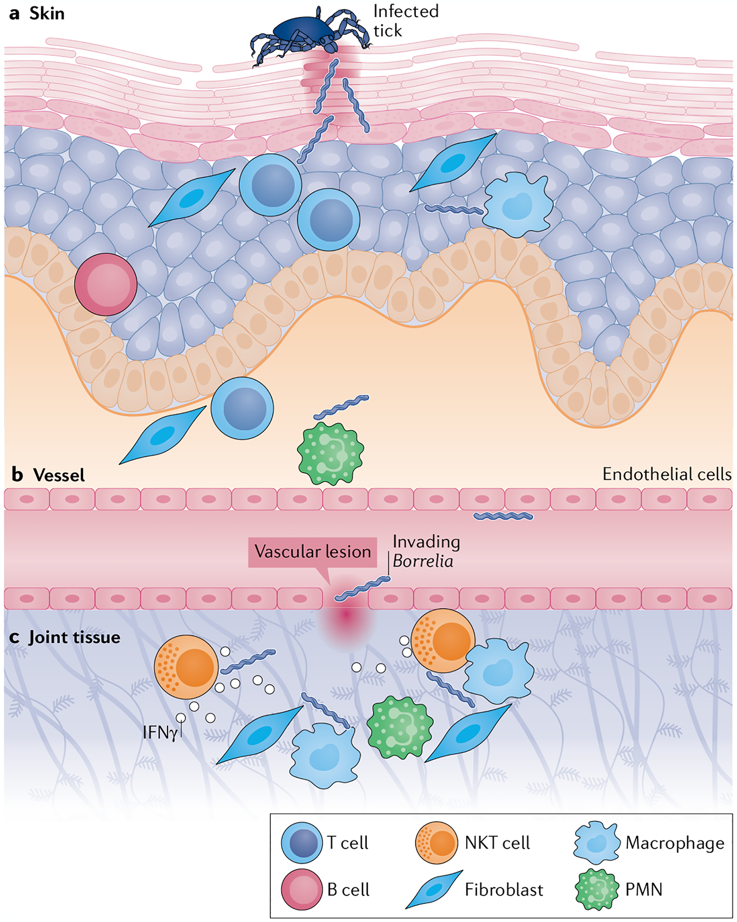
a | Spirochaetes invade the skin during the bloodmeal of an infected Ixodes tick. Upon entry, tissue-resident T cells, B cells, resident antigen-presenting cells (such as macrophages and dendritic cells), some polymorphonuclear cells (PMNs) and stromal cells (such as fibroblasts, keratinocytes, epithelial cells and endothelial cells) are responsible for early-acute immune responses to infection. b | A small number of spirochaetes escape the site of invasion and enter the vasculature, where Borrelia surface lipoproteins interact with vascular endothelial cells to induce the loosening of tight junctions. c | Spirochaetes enter the extracellular matrix of joint tissue through vascular lesions. Once in the joint tissue, they induce acute inflammatory responses by resident cells such as endothelial cells and synovial fibroblasts, which produce adhesion molecules, matrix metalloproteinases and innate immunity cytokines and chemokines. Natural killer T (NKT) cells produce IFNγ in response to CD1-presented immunogenic Borrelia glycolipids, thereby enhancing vascular barrier function and limiting spirochaetal invasion and chronic inflammation. The cytotoxic function of NKT cells might also directly contribute to spirochaetal killing. The nature and magnitude of these early immune responses in the skin and joint help to set the stage for subsequent arthritis development.