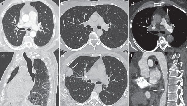
FIGURE 1.
Various imaging patterns of immunoglobulin G4-related disease thoracic involvement (each computed tomography (CT) scan section comes from a different patient). a) Axial contrast-enhanced thoracic CT scan showing an important bronchial thickening of the segmental right superior lobe bronchi (arrows). b) Axial unenhanced thoracic CT scan showing nodular ground–glass opacities in the posterior segment of the right superior lobe (arrows). c) Axial enhanced thoracic CT scan showing lymph nodes enlargement in the mediastinum in situation 5 and 10R (arrows). d) Sagittal reconstruction of thoracic CT scan showing distal bronchial distension and traction bronchiectasis in the interstitial lung pattern (circle). e) Axial unenhanced thoracic CT scan showing a mass in the right superior lobe (arrow). f) Sagittal enhanced aortic CT scan showing a stenosis of descending aorta and aortic wall thickening due to posterior mediastinitis (arrow).

