
Figure 5. Genetic and pharmacologic targeting of Galectin-3 protects against RGC loss in microbead-induced glaucoma.
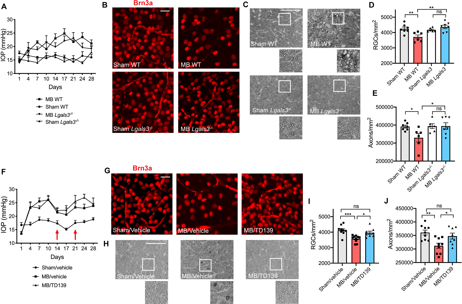
A) Intraocular pressure (IOP) measurements of microbead-injected (MB) and sham-injected wildtype (WT) and Lgals3−/− eyes (n=6–7 per group). B) Representative images of Brn3a+ retinal ganglion cells (RGCs) in MB and sham-injected WT and Lgals3−/− retinas. Scale bar 20 μm. C) Representative images of p-phenylenediamine (PPD) staining of optic nerves from MB and sham-injected WT and Lgals3−/− animals. Scale bar 50 μm. D) Quantification of RGC cell body numbers from (B) compared using one-way ANOVA. E) Quantification of axon counts from (C) compared using one-way ANOVA. F) IOP measurements of MB and sham-injected WT eyes treated with vehicle or Galectin-3 inhibitor TD139 (n=8–10 eyes per group). Intravitreal injections of the inhibitor or vehicle were administered at 2 and 3 weeks after MB injection (red arrows). G) Representative images of Brn3a+ RGCs in WT retinas treated with vehicle or TD139 inhibitor. Scale bar 20 μm. H) Representative images of PPD staining of optic nerves from WT animals treated with vehicle or TD139 inhibitor. Scale bar 50 μm. I-J) Quantification of RGC cell body numbers from (G) and axon counts from (H) compared using one-way ANOVA. Data in A-E and F-J were each pooled from two independent experiments. All results are shown as mean +/− SEM, *P<0.05, **P<0.01, ***P<0.001, ns = not significant.