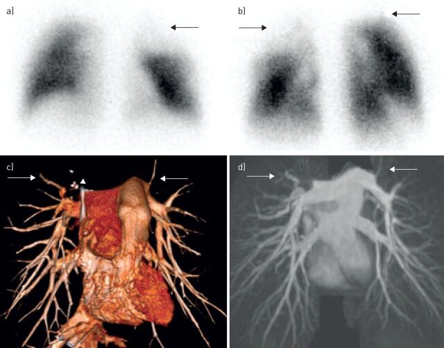
FIGURE 2.
A 53-year-old patient with fibrosing mediastinitis. a) Anterior and b) posterior views from a perfusion scintigraphy scan show multiple segmental perfusion defects (arrows). c) Volume-rendered computed tomography angiography and d) magnetic resonance pulmonary angiography demonstrate attenuated pulmonary vasculature (arrows) with calcified mediastinal lymph nodes (arrowhead).

