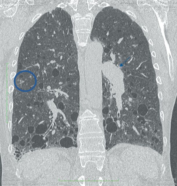
FIGURE 2.
A 63-year-old woman with Sjogren syndrome. Coronal computed tomography image shows numerous cysts admixed with fine reticular abnormalities in the lower lobes. In the upper lobes, a few centrilobular branching opacities (circle) consistent with bronchiolitis (likely follicular) can be appreciated. This patient also suffered from pulmonary hypertension, which is responsible for pulmonary artery enlargement (arrow).

