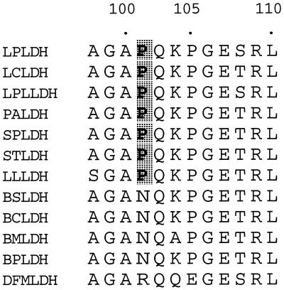
FIG. 3.
Alignment of amino acid sequences of the active-site loops of l-LDHs of bacilli and lactic acid bacteria. LPLDH, L. pentosus (28); LCLDH, L. casei (23); LPLLDH, L. plantarum (9); PALDH, Pediococcus acidilactici (12); SPLDH, Streptococcus mutans (30); STLDH, S. thermophilus (31); LLLDH, Lactococcus lactis (16, 24); BSLDH, B. stearothermophilus (2); BCLDH, B. caldotenax (3); BMLDH, B. megaterium (33); BPLDH, B. psychrosaccharolyticus (32); DFMLDH, dogfish muscle (31). Proline residues conserved in l-LDHs of lactic acid bacteria are shaded.

