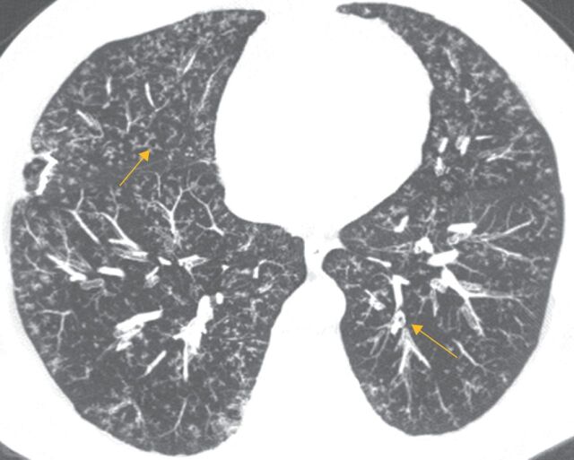
FIGURE 5.
Axial computed tomography scan demonstrating classic findings of follicular bronchiolitis in a 55-year-old male with rheumatoid arthritis. Note bilateral diffuse centrilobular peribronchial nodules <3 mm in size with branching structures corresponding to bronchial dilation and wall thickening (arrows). Image courtesy of Sudhakar Pipavath (University of Washington Medical Center, Seattle, WA, USA).

