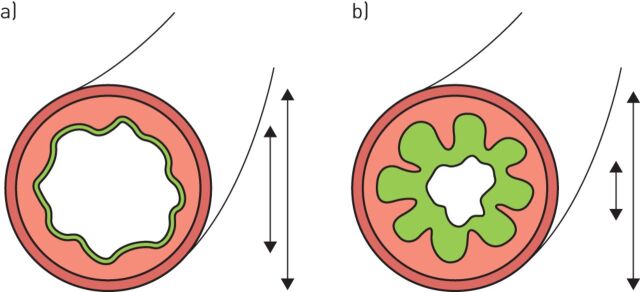
FIGURE 1.
a) A healthy small airway and b) an inflamed airway with a thickened wall at full inspiration. On inspiration, the mucosa of the healthy airway is only slightly folded. The inflamed airway has larger folds compared with the normal airway. Mucus fills up the gaps between the mucosal folds. On chest CT, the folds and mucus will be interpreted as a thickened airway wall. This figure also illustrates why the outer diameter is a more robust parameter for diagnosing and quantifying bronchiectasis because in contrast to the inner diameter, it is not influenced by the presence of mucus in the lumen. Image provided by, and reproduced and modified with the kind permission of, M. Meerburg (Amsterdam, The Netherlands).

