Abstract
Esophagitis dissecans superficialis (EDS) is an uncommon disease characterized by esophageal mucosal sloughing. EDS is a benign condition that usually resolves without residual pathology. Medication, chemical irritants, hot drinks, and autoimmune diseases have all been associated with EDS. Here a 60-year-old lady with post-COVID-19 EDS is presented. Her chief complaint was dysphagia and odynophagia for 2 weeks duration. EDS diagnosis was based on endoscopic findings and biopsy. Her problem was improved by a high dose of pantoprazole.
Keywords: EDS, COVID-19, Esophagitis dissecans superficialis
Introduction
In December 2019, the World Health Organization (WHO) was notified of idiopathic pneumonia in Wuhan, China. Investigations by Chinese scientists to illustrate the etiology led to the discovery of the Severe acute respiratory syndrome coronavirus 2 (SARS-CoV-2) infection in China.1 The first clinical findings reported were respiratory symptoms combined with bilateral pulmonary ground-glass lesions on CT scan and radiography.2 Thereafter, some extra pulmonary manifestations have also been reported as early disease manifestations or related complications, including gastrointestinal, hepatic, biliary, pancreatic, cardiovascular, ophthalmologic, and neurological manifestations.3,4 One of the rare gastrointestinal complications after SARS-CoV-2 infection may be esophagitis dissecans superficialis (EDS).
EDS is a desquamative disorder of the esophagus, with sloughing of the superficial mucosa. Endoscopically, EDS is characterized by sheets of sloughed squamous tissue with normal underlying mucosa.5 EDS can easily be misidentified or unrecognized due to its frequently vague presenting symptoms and gradual progression.6 This study reported an EDS case after COVID-19.
Case Report
A 68-year-old woman came to the clinic with the chief complaint of dysphagia to liquid and then solid material that gradually progressed, and odynophagia lasted for 2 weeks duration. She also had a history of anorexia, atypical chest pain, and 2-3 kg weight loss. One month earlier, the patient was admitted to the hospital for 10 days because of COVID-19. She received dexamethasone 8 mg/day and lopinavir/ritonavir (Kaletra) (400 mg and 100 mg, respectively), and heparin as prophylaxis, during hospital admission. Aspirin 80 mg/day was started for the patient after hospital discharge. The patient had no history of co-morbidities other than mitral valve prolapse. Personal history was also negative for cigarette smoking or alcohol consumption. Physical examination and primary para-clinical tests were within normal limits.
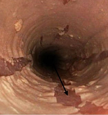
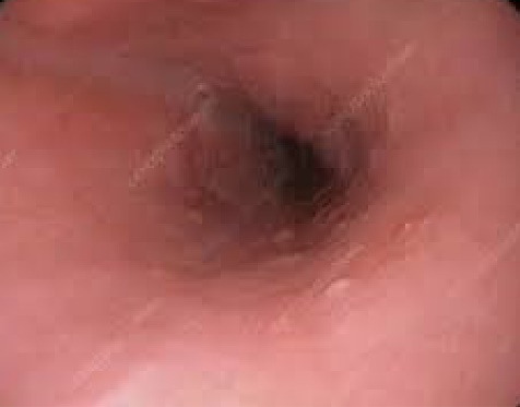
Endoscopy was performed that showed sloughing of the mucosal epithelium (Black arrow), the entire length of the esophagus, as a tubular cast within the esophageal lumen (Figure 1). Pathologic finding was in favor of esophageal squamous mucosal splitting of the superficial layer with minimal focal lymphocytic infiltration in the deepest layer with diffuse parakeratosis throughout (Figure 2). No neutrophilic and eosinophilic infiltrations were seen. No fungal or viral infections such as hepatitis B and cytomegalovirus were seen in microscopic and laboratory examinations, all in favor of EDS. She received a high-dose proton-pump inhibitor (PPI). Her symptoms gradually improved in one month, and her re-endoscopic assessment was normal. After re-evaluation was started, and in the follow-up after one month, it was suggested that her symptoms were healed. In the follow-up endoscopy, sloughed mucosa was healed (Figure 3).
Figure 1.

Esophagus endoscopic view (EDS).
Figure 2.

Biopsy of esophageal squamous mucosa: Diffuse separation of the superficial parakeratotic layer from the underlying epithelium (Black arrow). The basal layer is intact with a lack of significant degeneration or inflammation.
Figure 3.

Normal view after treatment.
Discussion
EDS is the term developed by Rosenberg in 1892 to describe an endoscopic finding characterized by the sloughing of large sections of epithelium, which can be coughed up or vomited up as an esophageal cast.7 Certain medications (bisphosphonates, non-steroidal anti-inflammatory drugs, potassium chloride), hot beverages, chemical irritants, celiac disease, collagen vascular disorders, and autoimmune bullous dermatoses (pemphigus and pemphigoid) have been associated with EDS, though the pathogenesis in most cases is unknown.7 In regard to the unknown nature of the SARS-CoV-2 accompanied with its variable complications, EDS can be attributed to the gastrointestinal side effects of COVID-19, although it may be due to Aspirin consumption.
Striped mucosa with or without bleeding, long linear mucosal breaks, vertical fissures, and circumferential cracks are all endoscopic characteristics of EDS.7 Sloughing and flaking of the superficial squamous epithelium with occasional bullous separation of the layers, parakeratosis, and variable degrees of acute or chronic inflammation are seen in biopsies from individuals with diagnosed EDS.8 EDS is usually a benign condition that resolves without lasting esophageal pathology, though have an acute dramatic presentation.7 Occasionally, the patient may present with obstructive symptoms due to esophageal casts.9 EDS is often confused with candida esophagitis, so it mandates pathological evaluation by taking a biopsy specimen, though candida may be seen in some cases without active inflammation.7,10
Management of EDS varies based on the patients’ severity of symptoms and underlying risk factors. Although in most cases, it resolves by self-loosening without any long-term side effects, PPIs may be effective, especially for further damage prevention. Immunosuppression has also been suggested in cases where the disease is likely to be autoimmune.6Due to the idiopathic cause of the disease in our patient, the patient was prescribed 40 mg of pantoprazole twice a day, and the patient was advised to be re-evaluated one month later. The patient’s symptoms improved gradually, and her esophageal EGD subsided without mucosal sloughing or stricture, documented by endoscopic and pathologic assessments.
Although non-documented cause and effect, this is the first case of EDS after COVID-19 infection. Based on review literature, there are less than 100 reported cases of EDS (and sloughing esophagitis) up to date although it seems to be more common. This may be due to vague self-limited symptoms or undiagnosed endoscopic or pathological findings.4
Please cite this paper as: Salehi AM, Salehi H, Hasanzarrini M. Esophagitis dissecans superficialis after COVID-19; a case report. Middle East J Dig Dis 2022;14(3):346-348. doi: 10.34172/mejdd.2022.293.
Footnotes
Conflict of Interest
The authors declare that there is no conflict of interest.
Ethical Approval
This study was approved by the Ethics Committee of Hamadan University of Medical Science (IR.UMSHA.REC.1400.262)
Funding/Support
This research received no specific grant from any funding agency in the public, commercial, or not-for-profit sectors
Authors’ Contribution
Study concept and design: AMS and HS. Acquisition of data: MH and HS. Critical revision of the manuscript for important intellectual content: AMS and HS. Administrative, technical, and material support: MH. Drafting of the manuscript: AMS and MH
References
- 1.Salehi AM, Salehi H, Mohammadi HA, Afsar J. SARS-CoV-2 and subacute thyroiditis: a case report and literature review. Case Rep Med. 2022;2022:6013523. doi: 10.1155/2022/6013523. [DOI] [PMC free article] [PubMed] [Google Scholar]
- 2.Zhou F, Yu T, Du R, Fan G, Liu Y, Liu Z. et al. Clinical course and risk factors for mortality of adult inpatients with COVID-19 in Wuhan, China: a retrospective cohort study. Lancet. 2020;395(10229):1054–62. doi: 10.1016/s0140-6736(20)30566-3. [DOI] [PMC free article] [PubMed] [Google Scholar]
- 3.Abobaker A, Raba AA, Alzwi A. Extrapulmonary and atypical clinical presentations of COVID-19. J Med Virol. 2020;92(11):2458–64. doi: 10.1002/jmv.26157. [DOI] [PMC free article] [PubMed] [Google Scholar]
- 4.Patel KP, Patel PA, Vunnam RR, Hewlett AT, Jain R, Jing R. et al. Gastrointestinal, hepatobiliary, and pancreatic manifestations of COVID-19. J Clin Virol. 2020;128:104386. doi: 10.1016/j.jcv.2020.104386. [DOI] [PMC free article] [PubMed] [Google Scholar]
- 5.Hart PA, Romano RC, Moreira RK, Ravi K, Sweetser S. Esophagitis dissecans superficialis: clinical, endoscopic, and histologic features. Dig Dis Sci. 2015;60(7):2049–57. doi: 10.1007/s10620-015-3590-3. [DOI] [PubMed] [Google Scholar]
- 6.Jaben I, Schatz R, Willner I. The clinical course and management of severe esophagitis dissecans superficialis: a case report. J Investig Med High Impact Case Rep. 2019;7:2324709619892726. doi: 10.1177/2324709619892726. [DOI] [PMC free article] [PubMed] [Google Scholar]
- 7.De S, Williams GS. Esophagitis dissecans superficialis: a case report and literature review. Can J Gastroenterol. 2013;27(10):563–4. doi: 10.1155/2013/923073. [DOI] [PMC free article] [PubMed] [Google Scholar]
- 8.Longman RS, Remotti H, Green PH. Esophagitis dissecans superficialis. GastrointestEndosc. 2011;74(2):403–4. doi: 10.1016/j.gie.2011.03.1117. [DOI] [PubMed] [Google Scholar]
- 9.Rokkam VR, Aggarwal A, Taleban S. Esophagitis dissecans superficialis: malign appearance of a benign pathology. Cureus. 2020;12(6):e8475. doi: 10.7759/cureus.8475. [DOI] [PMC free article] [PubMed] [Google Scholar]
- 10.Carmack SW, Vemulapalli R, Spechler SJ, Genta RM. Esophagitis dissecans superficialis (“sloughing esophagitis”): a clinicopathologic study of 12 cases. Am J Surg Pathol. 2009;33(12):1789–94. doi: 10.1097/PAS.0b013e3181b7ce21. [DOI] [PubMed] [Google Scholar]


