
Figure 4: IFI16/Ifi204 impacts osteoclast migration to the inflammatory-induced bone lesions.
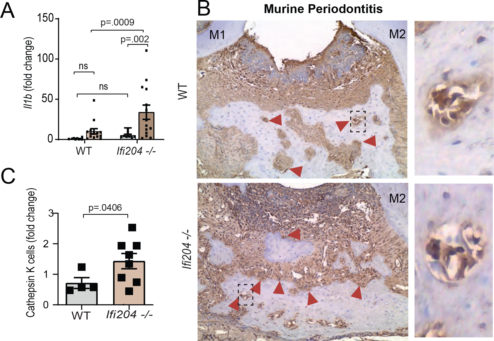
A) Il1b quantification in gingival tissues of WT and Ifi204−/− mice in health and 9d periodontitis. B) Red arrows indicate cathepsin K+ cells. Researchers were blind to group distribution and genotype when counting cathepsin K+ cells. Red arrows= cathepsin K+ cells. C) Cathepsin K histological staining of diseased periodontal tissues displayed a higher number bone-resorbing cells in Ifi204−/− mice (9d post-ligature placement) when compared to WT controls (magnification 20x).