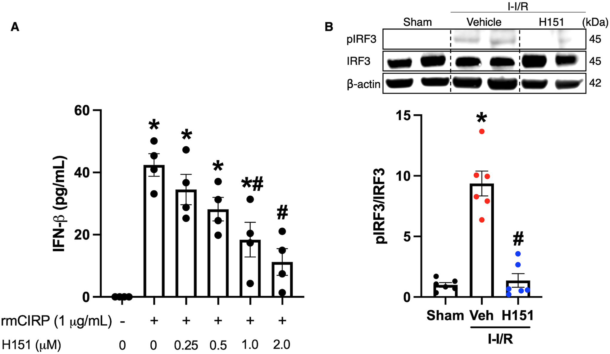
Figure 1. H151 inhibits STING activation in vitro and in vivo.

(A) RAW264.7 cells were pre-treated with H151 at various doses (0.25, 0.5, 1.0, 2.0 μM) one hour prior to stimulation with rmCIRP (1 μg/mL). Cells were then incubated for 24 hours, and culture supernatant was collected. IFN-β levels in the cell culture supernatant were measured using ELISA. Data are expressed as mean ± SEM and compared by ANOVA and SNK tests (*p < 0.05 vs control, #p < 0.05 vs rmCIRP alone; n = 4/group). (B) Intestines were collected from mice 4 hours after intestinal reperfusion and administration of intraperitoneal H151 or vehicle. Intestine tissue homogenates were assessed for pIRF3 and IRF3 proteins by Western blot. The blots were stripped and incubated with anti-β-actin antibodies to serve as the loading control. Representative Western blots for pIRF3, IRF3, and β-actin are shown. Each blot was quantified by densitometric analysis. pIRF3 expression in each sample was normalized to total IRF3 expression and the mean values of the sham group were standardized as 1 for comparison. Data are expressed as mean ± SEM and compared by the Kruskal-Wallis test (*p < 0.05 vs sham, #p < 0.05 vs vehicle, n = 6/group). I-I/R, intestinal-ischemia/reperfusion.
