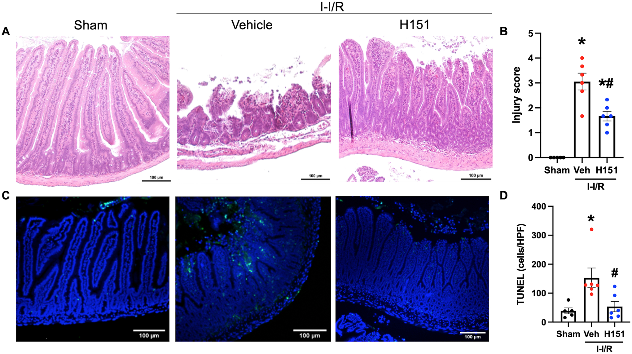
Figure 4. H151 reduces intestinal injury and cell death after intestinal I/R.

Organ injury was assessed by histological analysis using a validated scale of intestinal injury after I/R. (A) Representative images of H&E stained intestinal tissue at 100x (scale bar: 100μm). (B) Intestinal injury scores were calculated from zero to four with increasing score reflecting greater damage as assessed by blunting of the villi, decreased villi to crypt ratio, lymphocytic infiltration, and degree of necrosis (n = 5–6/group). TUNEL staining was performed to evaluate cell death. (C) Representative images of TUNEL stained sections at 100x (scale bar: 100μm). (D) TUNEL-positive cells were quantified using ImageJ software, and are expressed as cells/HPF (n = 5–6/group). Data are expressed as mean ± SEM and compared by (B) ANOVA and SNK tests, and (D) the Kruskal-Wallis test (*p < 0.05 vs sham, #p < 0.05 vs vehicle). I-I/R, intestinal-ischemia/reperfusion.
