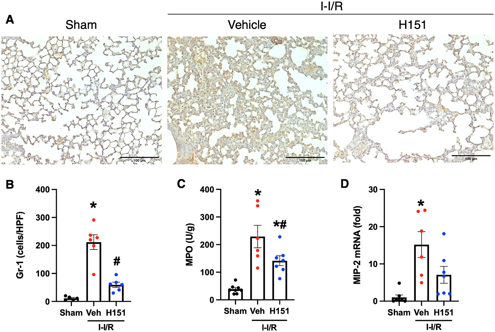
Figure 5. H151 reduces neutrophil infiltration and chemokine expression in lungs after intestinal I/R.

Four hours after reperfusion, lungs were collected. Immunohistochemical staining of Gr-1 neutrophils in lung tissue sections was performed. (A) Representative images of anti-Gr-1 immunohistochemical stains of lung tissue at are shown at 200x, (scale bar: 100μm). (B) Gr-1 neutrophil infiltration was quantified using ImageJ software (n = 5–6/group). (C) MPO activity in lung tissue was measured and is expressed in units per gram of protein (n = 7–8/group). (D) The mRNA levels of MIP-2 in lungs were measured by RT-qPCR, and normalized to β-actin (n = 6–7/group). Data are expressed as mean ± SEM and compared by ANOVA and SNK tests (*p < 0.05 vs sham, #p < 0.05 vs vehicle). I-I/R, intestinal-ischemia/reperfusion.
