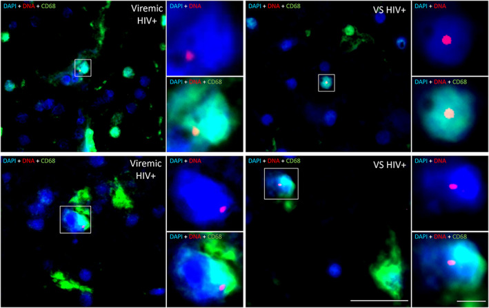
FIGURE 5.

CNS resident myeloid cells in virally suppressed PWH harbour HIV DNA. Representative images of HIV DNA+ CD68+ myeloid cells in frontal brain tissue from virally suppressed (VS HIV+; n = 2) or viremic PWH (n = 2) as determined by in situ hybridization for HIV DNA. HIV DNA (red), CD68 (green), or nuclei (blue) shown. Images acquired on a PALM Robo 4.2 at × 63 magnification. HIV DNA myeloid cells identified by white boxes. HIV DNA+DAPI+ and HIV DNA+CD68+DAPI+ insets of colocalization shown. Scale bars = 20 μm (×63 image) and 5 μm (×3.5 digital zoom ‐ insets). CNS = central nervous system; PWH = people with HIV; VS = virally suppressed.
