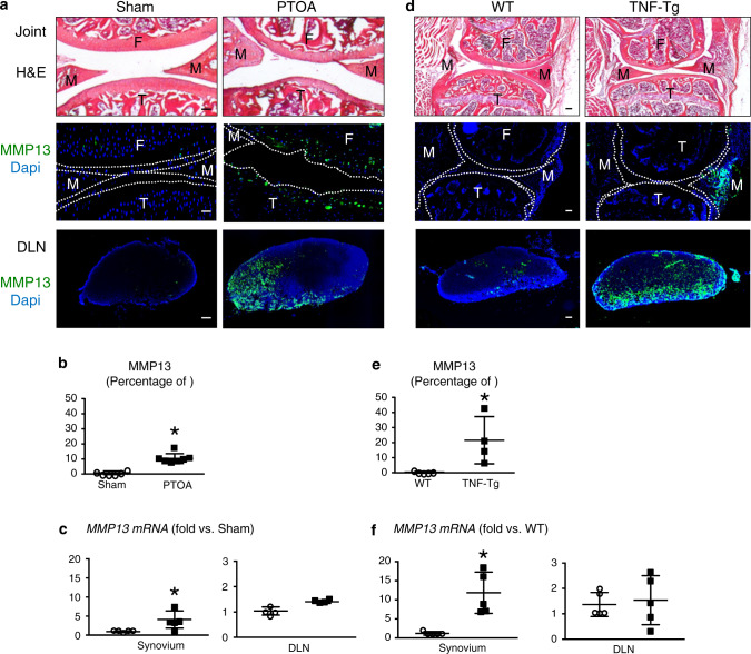
Fig. 6.
Arthritogenic MMP13 is removed from osteoarthritic and inflamed joints via the synovial lymphatic system. C57BL/6J mice received sham or DMM surgery (a–c), and TNF-Tg and WT control mice (d–f) were used. Knees and DLNs were harvested from operated mice 6 weeks post-surgery (a–c) and from 6-month-old WT and TNF-Tg mice (d–f). a, d Frozen sections from the knees were H&E stained for light microscopy, and representative 10x images are shown (scale bar = 100 µm). F: femur, T: tibia, M: meniscus. Parallel knee sections (dotted lines indicate the joint space) and DLN sections were immunostained with green fluorescent labeled antibodies against MMP13, counterstained with DAPI, and representative dark field images obtained at 10x are shown. Note that only articular chondrocytes in PTOA knees, synovium in TNF-Tg knees, and their DLNs, have robust MMP13 immuno-reactivity. b, e VisioPharm histomorphometry was performed to quantify the percentage of MMP13+ area of the DLN immunostained sections, and data are presented for each DLN +/− SD (*P < 0.05 vs. non-arthritic control with paired t-test). c, f Mmp13 mRNA levels in knees were assessed via qPCR. The data are presented for each tissue ± SD (*P < 0.05 vs. non-arthritic control with paired t-test). The content presented in Fig. 6 was derived from a PhD dissertation and was provided by Dr. Xi Lin, with her permission, a postdoctoral fellow in University of Rochester Medical Center258

