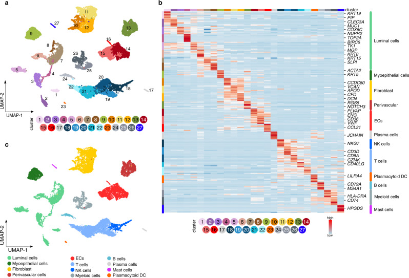
Fig. 3. Single cell taxonomy of the breast microenvironment.
a UMAP-plot of the subclustering of 18,082 cells from tumoral (n = 9) and matched peri-tumoral (n = 8) BC samples (TME and pTME respectively, composed of all cell types). b Heatmap of the expression levels of the top-10 marker genes in all 27 stromal subclusters derived from the non-EC enriched dataset (composed of all cell types). Color scale: red – high expression, blue – low expression. EC endothelial cell, DC dendritic cell, NK natural killer. c UMAP-plot of the same cells as in a, color-coded for the 12 major cell types.

