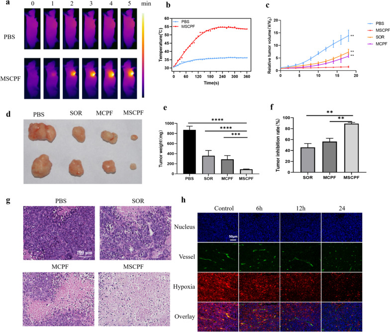
Fig. 7.
In vivo anti-tumor effect of MSCPF NPs. a IR thermal imaging by exposing tumors of mice to irradiation of 808 nm at 1.5 W cm−2 for 10 min after injection of PBS or MSCPF. b The temperature increase curve of tumors in the PBS and MSCPF groups after exposure to 808 nm irradiation. c Tumor growth curves of the mice during the experiment. d Photographs of the tumors harvested from mice at the end of treatment. e The weight of the tumors was measured. f The tumor inhibition rate of PBS, SOR, MCPF, or MSCPF on mice bearing SMMC-7721 tumors. g Representative HE staining histological sections of mouse tumors. h Immunofluorescence images of tumor slices of mice in control and the MSCPF-treated group after 6, 12, and 24 h injection. The nucleus, vessels, and hypoxic areas were respectively stained with 4′,6-diamidino-2-phenylindole (DAPI) (blue), anti-CD31 antibody (green), and anti-pimonidazole antibody (red)

