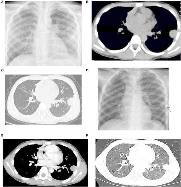
Figure 1.
Radiological images before ultransonography (US)-guided lung biopsy. (A) Signs of infiltration present in the left lower lung field. (B,C) Plane computed tomography (CT) at the previous hospital revealing a round soft tissue mass and a small amount of pleural effusion in the left lung. (D) Chest X-ray revealed a left lung nodule measuring 2 cm in diameter. (E,F) Contrast-enhanced CT of the chest showing a rim-enhancing lesion in the left lower lobe lung.

