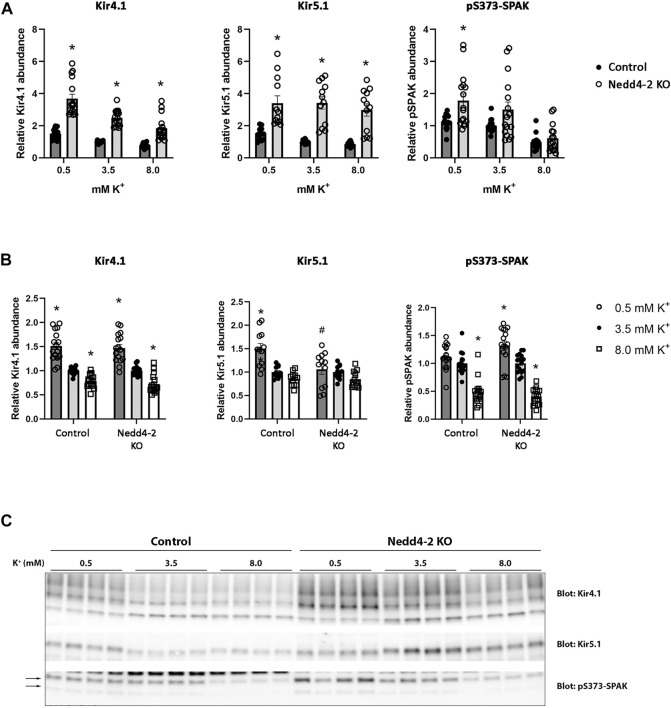
FIGURE 9.
Kir4.1, Kir5.1, and pS373-SPAK levels are increased in tubule suspensions isolated from Nedd4-2 KO mice.(A+B) Semi-quantitative assessment of Kir4.1, Kir5.1, and pS373-SPAK levels in tubules isolated from control and Nedd4-2 KO mice treated with 0.5 mM, 3.5 mM, or 8.0 mM K+ for 24 h. (A) Data are means ± S.E.M and normalized to control tubules treated with 3.5 mM K+. Difference between control and KO tubules treated with either 0.5 mM, 3.5 mM, or 8.0 mM K+ is assessed by 2way ANOVA followed by Tukey’s multiple comparisons test (*p < 0.05). (B) Data are means ± S.E.M. Control and KO tubules are normalized to their own 3.5 mM K+ group. Differences of Kir4.1, Kir5.1, and pS373-SPAK levels with different K+ concentrations in either control or KO tubules are assessed by 2way ANOVA followed by Tukey’s multiple comparisons test (*p < 0.05). Fold changes between control and KO tubules with changing K+ concentrations are assessed in the same 2way ANOVA analysis (# p < 0.05) (n = 12–16). (C) Representative immunoblots of Kir4.1, Kir5.1, and pS373-SPAK in control and KO tubules treated with 0.5 mM, 3.5 mM, or 8.0 mM K+.

