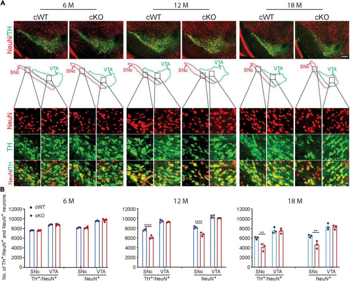
FIGURE 1.
Neurodegeneration in 12- and 18-month-old Pitx3cKO mice. (A) IFC co-staining of TH and NeuN in the ventral midbrain sections from 6-, 12-, and 18-month-old Pitx3cWT and Pitx3cKO mice. SNc and VTA were outlined, respectively (scale bar: 200 μm; high-magnification, 20 μm). (B) Quantification of TH+/NeuN+ and NeuN+ neurons in the SNc and VTA from 6-, 12-, and 18-month-old Pitx3cWT and Pitx3cKO mice (N = 3–4 mice per genotype; all males except for two females in 6-month-old Pitx3cKO and 12-month-old Pitx3cWT). 2way ANOVA analysis with Sidak’s multiple comparisons test, ****p < 0.0001 (12 months for TH+/NeuN+ co-staining), ****p < 0.0001 (12 months for NeuN+ staining), **p = 0.0041 (18 months for TH+/NeuN+ co-staining), **p = 0.0064 (18 months for NeuN+ staining).

