Figure 1.
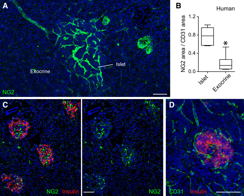
Blood vessels in human pancreatic islets are densely covered by pericytes. A: Maximal projection of confocal images of a human pancreatic section immunostained for the pericyte marker NG2 (green). The majority of NG2-expressing cells is present in islets in the human pancreas. NG2 is also expressed by smooth muscle cells around arteries and islet-feeding arterioles. B: Quantification of NG2 immunostained area normalized to CD31 immunostained area in endocrine (islets) and exocrine tissue compartments of the human pancreas. Data are shown in a box and whisker plot (min to max) (n = 31 islets from four different donors without diabetes). *P < 0.05 by unpaired t test. C: Confocal images of a human pancreatic section immunostained for insulin (red) and NG2 (green). Regions rich in NG2-expressing cells are islets. D: Maximal projection of confocal images of a human pancreatic section immunostained for the endothelial cell marker CD31 (green) and insulin (red). Scale bars = 50 μm (A, C, and D).

