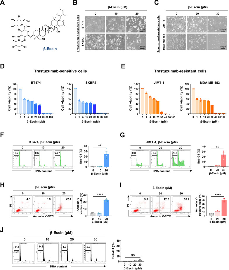
Fig. 1.
β-escin induces apoptosis in trastuzumab-sensitive and –resistant cells. A Chemical structure of β-escin. B The changes in morphology of BT474 and SKBR3 cells after treatment of β-escin (10-20 μM, 48 h) as observed by phase-contrast microscopy. C Representative phase-contrast images of JIMT-1 and MDA-MB-453 cells after treatment with β-escin (10-30 μM, 48 h). D, E Trastuzumab-sensitive SKBR3 and BT474 cells (D) and trastuzumab-resistant MDA-MB-453 and JIMT-1 cells (E) were treated with various concentrations of β-escin (1-100 μM) for 48 h, and cell viability was evaluated by MTS assay (****p<0.0001). F, G BT474 (F) and JIMT-1 cells (G) were treated with β-escin (10-20 μM and 20-30 μM, respectively) for 48 h, and the percentages of cells in the sub-G1 phase were quantified using flow cytometry (**p<0.01). H, I The percentages of the early and late apoptotic cells in BT474 (H) and JIMT-1 cells (I) following exposure to β-escin (10-20 μM and 20-30 μM, respectively) were determined by annexin V/PI staining (****p<0.0001). J Normal human mammary gland epithelial MCF10A cells were treated with β-escin (10-30 μM) for 48 h, and the sub-G1 fraction was analyzed (not significant, NS). The results are expressed as mean ± SEM after three independent experiments and analyzed by one-way ANOVA and Bonferroni’s post hoc test

