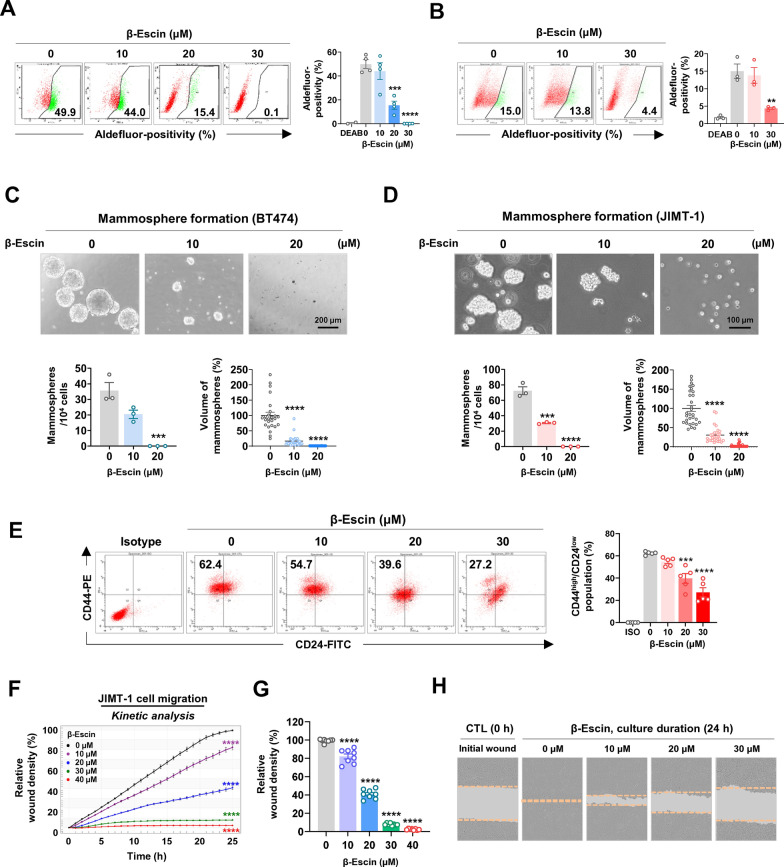
Fig. 4.
β-escin impairs CSC-like properties. A, B BT474 and JIMT-1 cells were treated with β-escin (10-30 μM) for 48 h and ALDH1 activity was assessed by flow cytometry. DEAB was used to define the baseline of Aldefluor-positive fluorescence. The quantitative graphs represent the percentage of Aldefluor-positive cells in BT474 (A, ***p<0.001) and JIMT-1 cells (B, **p<0.01). C, D BT474 (C, 5×10⁴ cells/ml) and JIMT-1 (D, 1.5×10⁴ cells/ml) were treated with β-escin (10-20 μM) in serum-free suspension conditions for 4 and 8 days, respectively. The number and volume of mammospheres were significantly reduced in the presence of β-escin, and the quantitative graphs are shown in the bottom panels, respectively (***p<0.001). E Effect of β-escin (10-30 μM, 48 h) on the CD44high/CD24low stem-like phenotype in JIMT-1 cells. CD44high/CD24low populations were determined by flow cytometry and were quantified (right panel, ***p<0.001). F JIMT-1 cells were treated with β-escin (0-40 μM) for 24 h. Cell migration was kinetically monitored with an IncuCyte™ System and quantified for the indicated time durations (****p<0.0001). G The quantitative graph represents the percentages of relative wound density of JIMT-1 cells at 24 h (****p<0.0001). H Representative images of wound closure by cell migration at 0 and 24 h after β-escin treatment (0-30 μM). The orange dotted line indicates the edge of the scratched wound

