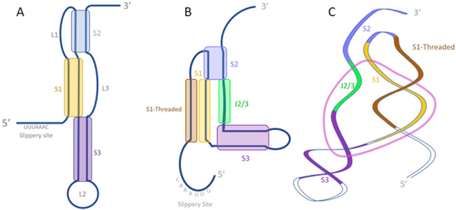
FIG 7.
Differences in structure prediction for the SARS-CoV-2 frameshifting pseudoknot. (A) Secondary structure described by Plant et al. (174) using 2-dimensional nuclear magnetic resonance (NMR). (B) Secondary structure described by Zhang et al. (178) using SHAPE. (C) Tertiary structure described by Zhang et al. using cryo-EM map guided modeling. For panels B and C, the dark brown highlighted segment represents the stem strand described as “threaded” through the” ring” (displayed in light pink) formed by the other strand of stem 1 (light brown), stem 2 (blue), stem 3 (purple), and an unpaired region between stem 2 and stem 3 (J2/3) (green).

