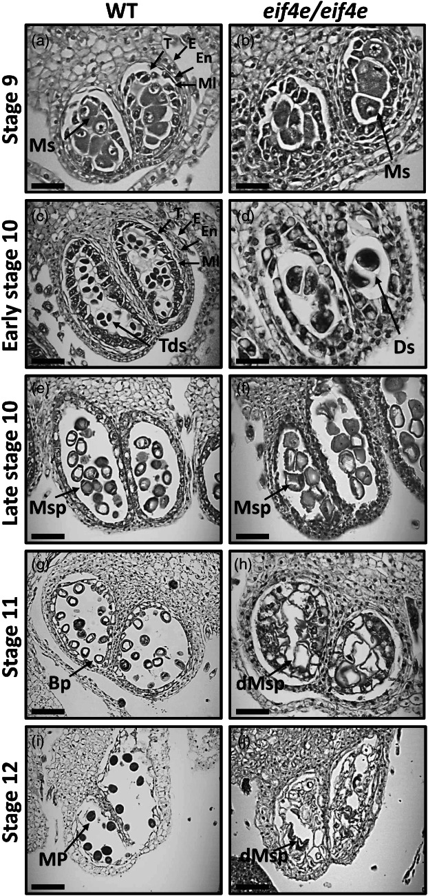
Figure 4.

Transversal sections of anthers throughout development in wild‐type (WT) and eif4e mutant (eif4e/eif4e) observed by light microscopy. Locules from the WT (a, c, e, g, i) and eif4e mutant (b, d, f, h, j) anthers from stages 9 to 12 of development. BP, bicellular pollen; dMsp, degraded microspores; Ds, dyads; E, epidermis; En, endothecium; ML, middle layer; MP, mature pollen; Ms, microsporocyte; Msp, microspores; T, tapetum; Tds, tetrads. Scale bars = 50 μm (a, b, c, d, e, f), 100 μm (g, h, i, j).
