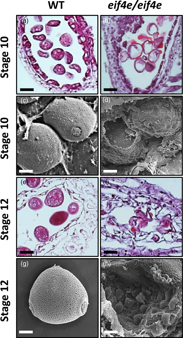
Figure 5.

Light microscopy (a, b, e, f) and scanning electron microscopy (c, d, g, h) images of transversal sections throughout anther development in the wild‐type (WT) and eif4e mutant (eif4e/eif4e). (b, d) Locules are filled by large amounts of stained/dark material that could correspond to sporopollenin in late stages 10. (f, h) Locules showing clumping of microspores unstructured as well as other stained/dark material of unknown origin in early stage 12. Scale bars = 25 μm, (a, b, e, f), 10 μm (c, d), 5 μm (g, h). [Colour figure can be viewed at wileyonlinelibrary.com]
