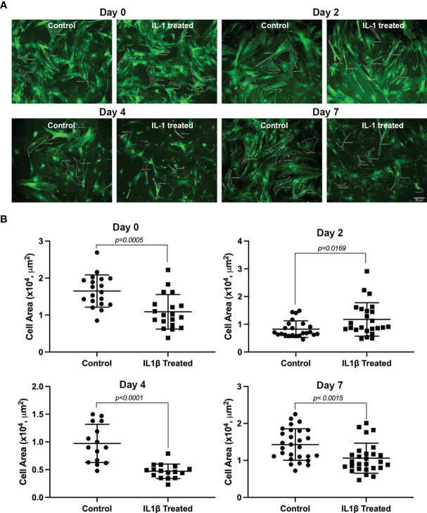
Figure 2.
Effect of IL-1β on MEC size. Lacrimal gland MEC were either left untreated (control) of incubated with IL-1β (10 ng/ml) for 2, 4, or 7 days. Three or 4 random images were taken, using the same camera setting for all conditions, from each well MEC size was quantified using SPOT Imaging software, as described in the Methods section. (A) Shows representative images from control and treated MEC at all time points measured and (B) Shows averaged data from 4 independent experiments. Compared to the control group, IL-1β treatment significantly decreased MEC size at all time points measured (Student’s t-test). Data are means ± SD; n=23-24 for day 0; n=19 for day 2; n=15-16 for day 4; and n=27-28 for day 7 with all data from 4 independent experiments. Scale bar = 50 μm.

