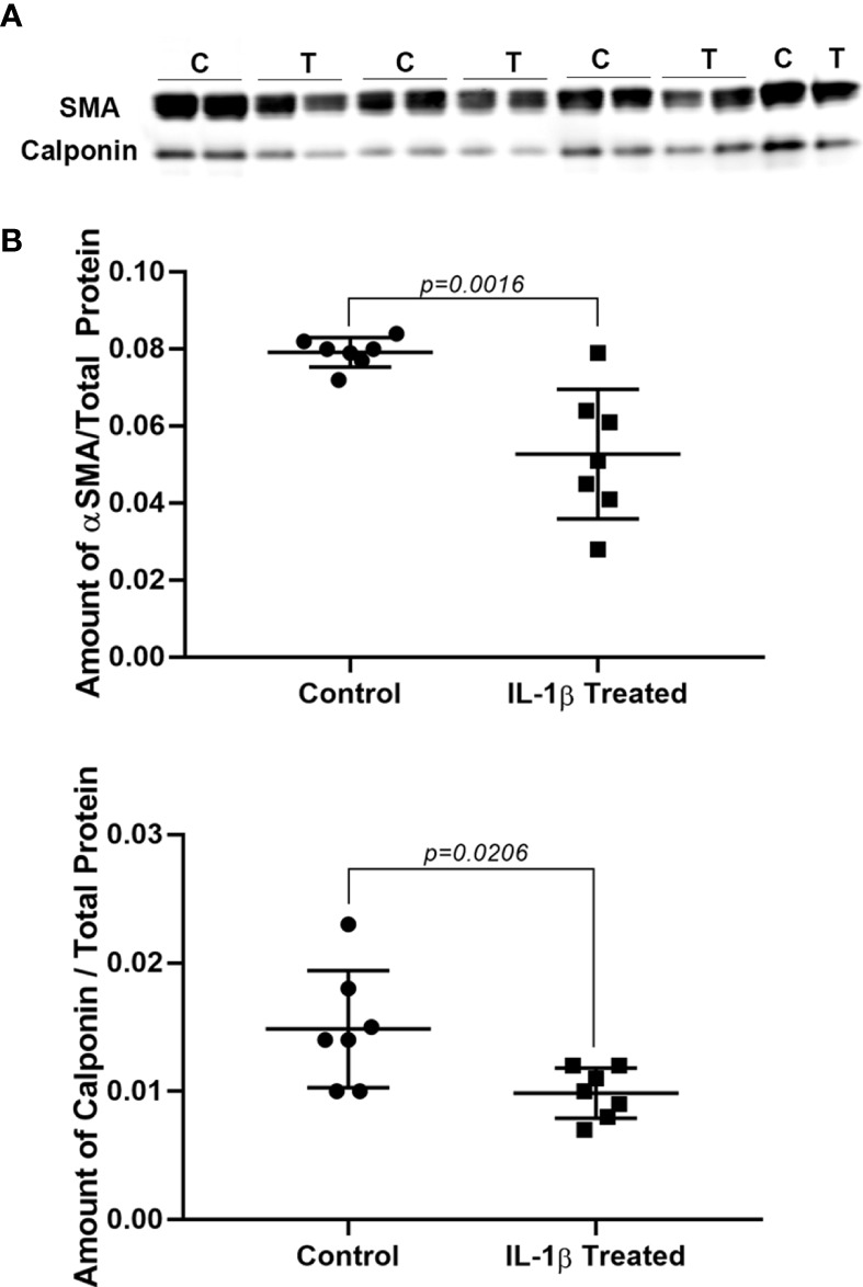
Figure 3.

Effect of IL-1β on SMA and calponin protein levels. Lacrimal gland MEC were either left untreated (control) or treated with IL-1β (10 ng/ml) for 7 days. SMA and calponin protein level were quantified by western blotting and reported as a ration relative to total protein stain, as described in the Methods section. (A) Shows western blots for SMA (top) and calponin (bottom) in control (B) and IL-1β treated (T) MEC samples. (B) Graphs showing the amount of SMA and calponin in control and treated lacrimal gland MEC relative to total protein stain. SMA and calponin amounts are significantly decreased in lacrimal gland MEC treated with IL-1β compared with the control (P = 0.0016 and P = 0.0206; respectively, Student’s t-test.). Data in the plots are means ± SD, n = 7 from 4 independent experiments.
