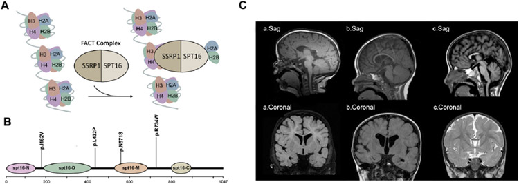
Figure 1.
Molecular and clinical findings in patients with SUPT16H mutations. (A) A schematic model of the FACT complex interacting with the nucleosome. (B) Diagram of Spt16 protein indicating the location of principal domains and the variants identified. The mutations are grouped according to their locations on Spt16 domains. (C) Callosal abnormalities in selected patients with SUPT16H changes: (a) T1-weighted sagittal and coronal images demonstrating a thin corpus callosum and diminished white matter volume in patient 1; (b) T1-weighted sagittal and coronal images demonstrating a thin corpus callosum and enlarged lateral ventricles in patient 4; (c) T1 sagittal and T2 FLAIR coronal images demonstrating partial agenesis of the corpus callosum in patient 5. FACT, facilitates chromatin transcription; FLAIR, fluid-attenuated inversion recovery.

